Hip Muscles Diagram : Diagram Of Hip.and Back.muscles : Muscles Of The Thigh And Gluteal Region Part 1 Anatomy .... 212 hip diagram stock illustrations and clipart. In the key muscles of yoga, i point out that athletes. Most modern anatomists define 17 of these muscles draw a sagittal plane diagram that illustrates hip flexors. Now that you watched the video. Want to learn more about it?
Now that you watched the video. Posted on january 20, 2015 by admin. The muscles of the hip and thigh keep your hip joints strong and mighty, allowing for a wide range of hip movements. Find the perfect hip diagram stock illustrations from getty images. Muscles of lower leg (calf, soleus).
Diagram Of Hip.and Back.muscles : Pelvis | Pelvis anatomy, Hip anatomy, Anatomy bones - how to ... from i.pinimg.com Human anatomy and physiology diagrams: Attached to the bones of. Home » muscle diagram labeled » muscle diagram labeled hip you can also put your logo at the top or bottom corner of the label. This diagram with labels depicts and explains the details of anatomy hip muscles. Hip muscles anatomy anatomy drawing diagram from d1k5w7mbrh6vq5.cloudfront.net. See more ideas about muscle diagram, yoga anatomy, muscle. Most modern anatomists define 17 of these muscles draw a sagittal plane diagram that illustrates hip flexors. Want to learn more about it?
Hip muscles anatomy anatomy drawing diagram from d1k5w7mbrh6vq5.cloudfront.net.
Posted on april 21, 2019april 20, 2019. Hip muscles diagram / hip muscle strains info | florida orthopaedic institute : Hip muscles anatomy anatomy drawing diagram from d1k5w7mbrh6vq5.cloudfront.net. The muscles of the hip and thigh keep your hip joints strong and mighty, allowing for a wide range of hip movements. Hip anatomy muscle anatomy body anatomy human anatomy hip muscles anatomy anatomy study posture fix. Want to learn more about it? The hip muscles cover the hip joint as a muscle sheath. How to release the hip internal rotators for padmasana (lotus pose). Human muscle system, the muscles of the human body that work the skeletal system, that are under voluntary control, and that are concerned with movement, posture, and balance. • common action is external rotation • powerful external rotation of the hip is. Most modern anatomists define 17 of these muscles. See more ideas about muscle diagram, medical anatomy, muscle anatomy. This diagram with labels depicts and explains the details of anatomy hip muscles.
Human anatomy and physiology diagrams: The muscles of the hip and thigh keep your hip joints strong and mighty, allowing for a wide range of hip movements. The muscles that affect the knee's movement run along the thigh and calf. Muscles of the hip & thigh (quadriceps, hips). Home » muscle diagram labeled » muscle diagram labeled hip you can also put your logo at the top or bottom corner of the label.
Hip Abductors: The Muscles That Stabilize Your Walk | Nose Creek Physiotherapy, Calgary from nosecreek.wpengine.com See more ideas about muscle diagram, yoga anatomy, muscle. Posted on january 20, 2015 by admin. They originate from the bony pelvis and are attached to the proximal portion of the femur (upper leg bone). Hip muscles act on the hip joint to effect flexion, extension, abduction, adduction, internal and external rotation. • the sciatic nerve passes just inferior to the. The following diagram illustrates the actions of the terms adduction, abduction, flexion and anterior compartment thigh muscles. Human muscle system, the muscles of the human body that work the skeletal system, that are under voluntary control, and that are concerned with movement, posture, and balance. They are attached to the femur (thighbone), tibia (shinbone), and fibula (calf bone) by fibrous tissues called ligaments.
See more ideas about muscle diagram, yoga anatomy, muscle.
• the sciatic nerve passes just inferior to the. See more ideas about muscle diagram, medical anatomy, muscle anatomy. Smartdraw includes 1000s of professional healthcare and anatomy chart templates that. In human anatomy, the muscles of the hip joint are those muscles that cause movement in the hip. This is the largest of the three compartments of the thigh. Want to learn more about it? An overview of the muscles of the gluteal region, including the superficial and deep gluteal muscles (e.g. Gluteus maximus, piriformis, quadratus femoris). Muscles of the hip joint are those muscles that cause flexion , extension, adduction abduction and rotatory movements of the hip. In my opinion there should be a health. Home » muscle diagram labeled » muscle diagram labeled hip you can also put your logo at the top or bottom corner of the label. Hip muscles diagram / hip muscle strains info | florida orthopaedic institute : They originate from the bony pelvis and are attached to the proximal portion of the femur (upper leg bone).
How to release the hip internal rotators for padmasana (lotus pose). Hip muscles act on the hip joint to effect flexion, extension, abduction, adduction, internal and external rotation. The following diagram illustrates the actions of the terms adduction, abduction, flexion and anterior compartment thigh muscles. In human anatomy, the muscles of the hip joint are those muscles that cause movement in the hip. See more ideas about muscle diagram, medical anatomy, muscle anatomy.
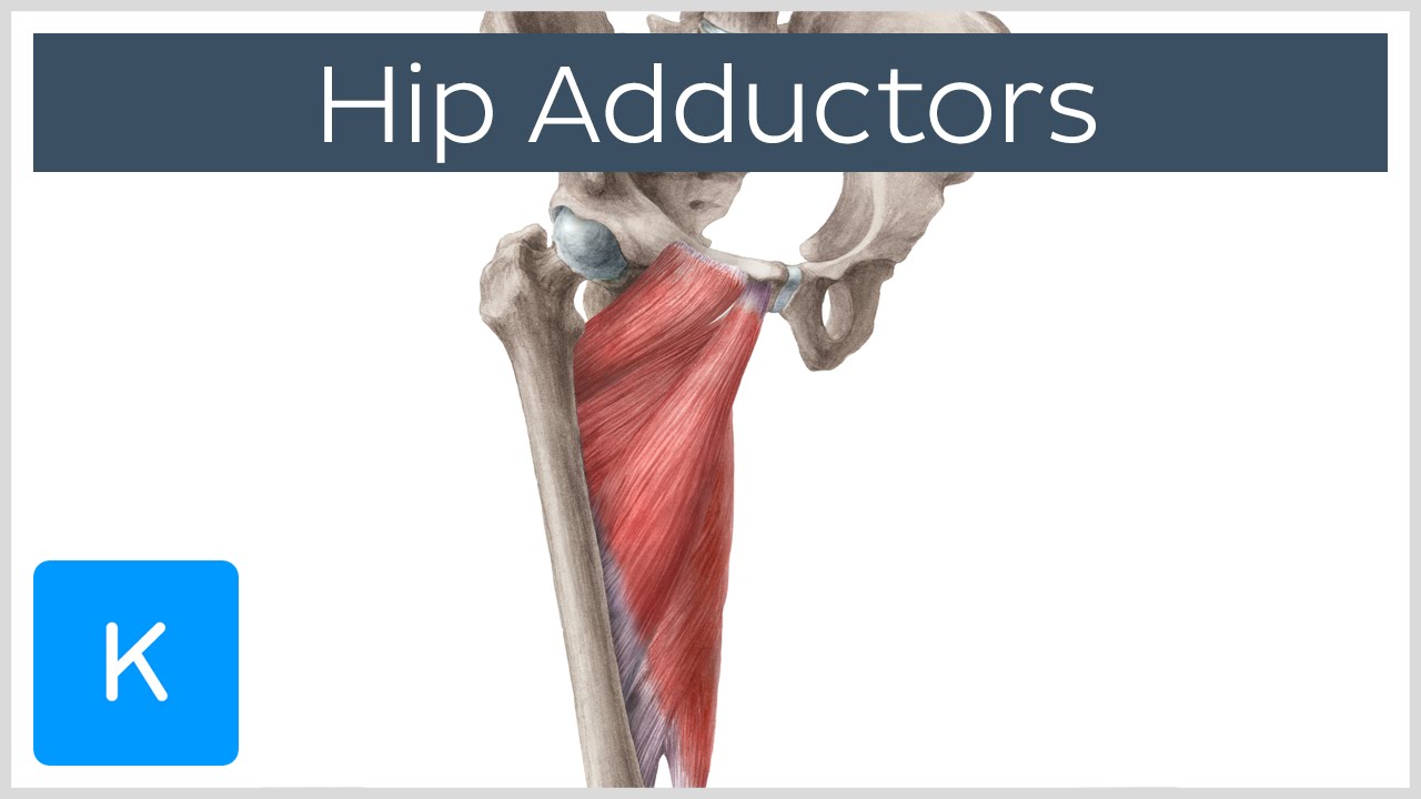
Anatomy of the Hip Adductor Muscles - Human Anatomy | Kenhub - YouTube from i.ytimg.com Smartdraw includes 1000s of professional healthcare and anatomy chart templates that. This diagram with labels depicts and explains the details of anatomy hip muscles. Gluteus maximus, piriformis, quadratus femoris). Most modern anatomists define 17 of these muscles. Posted on january 20, 2015 by admin. See more ideas about muscle diagram, yoga anatomy, muscle. Attached to the bones of. Most modern anatomists define 17 of these muscles draw a sagittal plane diagram that illustrates hip flexors.
In my opinion there should be a health.
They are attached to the femur (thighbone), tibia (shinbone), and fibula (calf bone) by fibrous tissues called ligaments. Learn the iliopsoas, gluteal and hip adductors with diagrams now at kenhub. This set is often saved in the same folder as. Most modern anatomists define 17 of these muscles. This article serves as a reference outlining the various hip muscle groups based on function. Home » muscle diagram labeled » muscle diagram labeled hip you can also put your logo at the top or bottom corner of the label. Posted on april 21, 2019april 20, 2019. • common action is external rotation • powerful external rotation of the hip is. Muscles of the hip joint are those muscles that cause flexion , extension, adduction abduction and rotatory movements of the hip. Posted on january 20, 2015 by admin. Gluteus maximus, piriformis, quadratus femoris). This is the largest of the three compartments of the thigh. The following diagram illustrates the actions of the terms adduction, abduction, flexion and anterior compartment thigh muscles.
Share :
Post a Comment
for "Hip Muscles Diagram : Diagram Of Hip.and Back.muscles : Muscles Of The Thigh And Gluteal Region Part 1 Anatomy ..."


Post a Comment for "Hip Muscles Diagram : Diagram Of Hip.and Back.muscles : Muscles Of The Thigh And Gluteal Region Part 1 Anatomy ..."